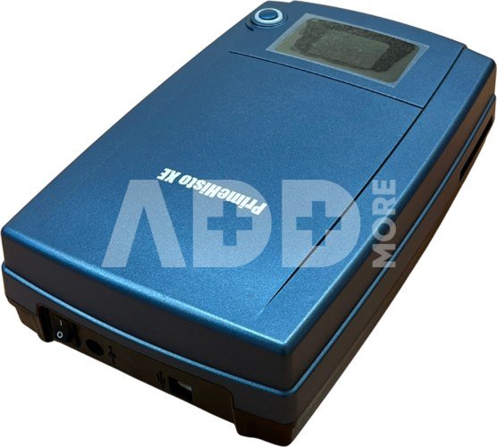
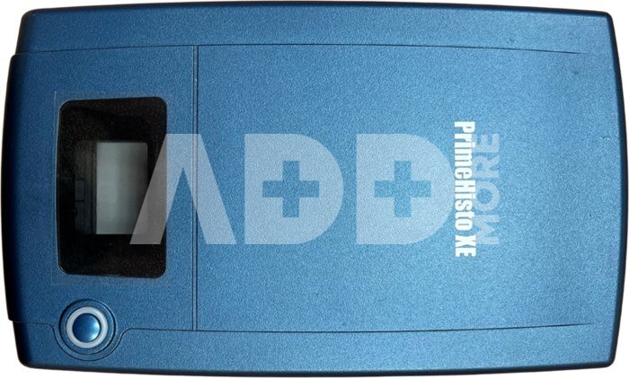
Pacific Image PrimeHisto XE
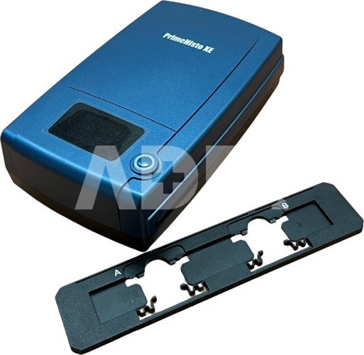
The PrimeHisto XE bridges the gap between traditional microscopy and digital workflows. It offers unmatched detail, real color accuracy, and powerful software tools in a compact, reliable package. Whether you’re documenting delicate histological preparations or building a digital archive for teaching and review, this scanner is optimized to deliver the clarity and consistency your work demands.Key Features & Benefits10,000 dpi resolutionCapture fine structural details in your tissue slides. Every cellular boundary, stain gradient, and morphological nuance is recorded with sharpness and accuracy. (Pacific Image Electronics Co., Ltd)True color reproduction with a linear CCD sensorUnlike area sensors that interpolate color, the PrimeHisto XE uses a True Color Linear CCD for direct RGB capture, preserving the actual stain colors and intensities in your slides. (Pacific Image Electronics Co., Ltd)Stable slide holding systemIncludes a dual-slide holder with secure alignment strips, ensuring that slides remain firmly positioned during scanning. (Pacific Image Electronics Co., Ltd)Broad scanning range to support microscope integrationThe scanner allows observation from about 10 µm up to 1 cm, making it easier to locate regions of interest and integrate with your microscope workflows. (Pacific Image Electronics Co., Ltd)Rich software support with HistoViewBundled with Pacific Image’s HistoView software, you gain access to features like gamma curve adjustment, fine control over brightness, contrast, saturation, and more—designed specifically for histological image processing. (Pacific Image Electronics Co., Ltd)Flexible compatibilityWorks on both Windows (7 through 11) and macOS 10.13+ systems. (Pacific Image Electronics Co., Ltd)USB 2.0 connectivity, with recommended host hardware of minimum 4 GB RAM (8 GB preferred). (Pacific Image Electronics Co., Ltd)SpecificationsScanning MediaMicroscope slideResolutionUp to 10,000 dpiDynamic Range3.9Light SourceWhite LED (transmission)SensorLinear array color CCDData Mode48-bit (color), 16-bit (grayscale)Scan Area24.3 mm × 36.5 mmFile FormatsTIFF, BMP, JPEGPower SupplyAC 100–240 V → 12 V DC, 1.5 ADimensions (L×W×H)Approx. 275 × 167 × 80 mmWeight2.1 kgOS SupportWindows 7/8/10/11, macOS 10.13+Recommended Requirements≥4 GB RAM, 50 GB free disk space (Pacific Image Electronics Co., Ltd)Ideal Use CasesAcademic and research labs conducting histological or pathological examinationsMedical / clinical labs requiring digital archiving of slidesTeaching institutions where students learn tissue morphology and stainingAny environment needing high-fidelity digitization of microscope slides
Discuss with a consultant in Kaunas
We don't have
Klaipėda Address: Birželio 23-iosios g. 23g Working hours: I-V 09:00 - 18:00 VI-VII not working Phone: +370 674 59488
Discuss with a consultant in Klaipėda
We don't have
Klaipėda Address: Taikos pr. 17 Working hours: I-V 10:00 - 19:00 VI 10:00 - 14:00 VII not working Phone: +370 604 43972


Reviews
0 average rating (0 votes)